
Case: 9 Scapulothoracic Crepitus
Crepitus around the scapulothoracic region maybe due to a predisposing abnormal anatomy such as,
- Malunion of scapula or rib fractures
- A history of resection of 1st rib for thoracic outlet syndrome
- Superomedial hooking of the scapula – approximately 6% of scapulae
It may also be due to,
- Overuse with normal anatomy
- Inflammation secondary to overuse causing bursal fibrosis
- Bony and / or soft tissue masses such as osteochondroma, elastofibroma dorsi, or scapular chondrosarcoma
In this case a 42 year old female patient presented with a 1-year history of discomfort and ‘clicking’ around the left non-dominant scapula. Onset had been insidious. Problems were described as being related to activity. No other medical problems were reported.
Examination demonstrated a full range of both shoulder and thoracic spine movements. A palpable crepitus and intermittent swelling was felt when the hand was placed over the left scapula and the patient asked to move their arm.
An X-ray demonstrated no bony abnormalities around the scapula or ribs.
An ultrasound demonstrated an echogenic swelling which could be seen extending from below the inferomedial border of the scapula with the arm placed in horizontal adduction across the chest. The full report reads:
Ultrasound of the inferomedial aspect of the left scapula demonstrated a heterogeneous swelling which appeared to lie deep to the subcutaneous tissue and superficial fascia measuring approximately 5 x 5 x 1.5 cm. The swelling contained no peripheral or internal vascularity. The swelling appeared to extend from below the inferior border of the scapula.
Clinically the patient described the swelling as having been present for approximately 1-year. No other medical issues were described. The swelling was non-tender to palpation.
Ultrasound findings are relatively nonspecific but do not have the typical appearance of a lipoma. Given appearance and location the swelling could represent an elastofibroma dorsi.
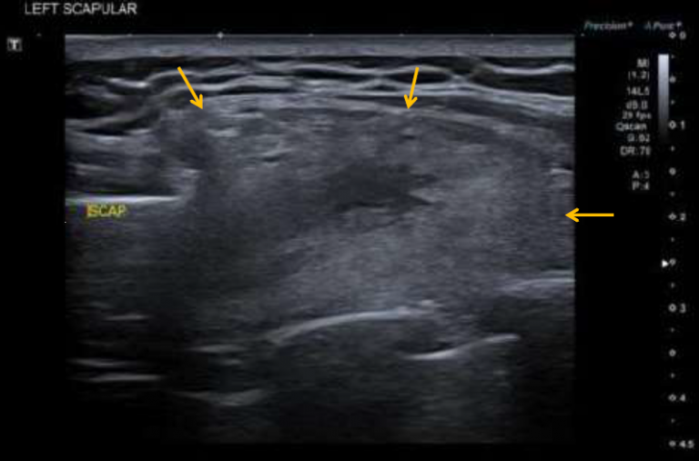
The inferomedial border of the scapula maybe seen to the left of this image. Extending from below the scapula maybe seen an echogenic swelling (yellow arrows). The ribs are seen laying below the swelling
The patient was referred to Orthopaedics and following surgery the diagnosis of an elastofibroma dorsi was confirmed.
Elastofibroma dorsi
Elastofibroma dorsi is a benign soft-tissue tumour with a characteristic location and imaging appearance. It is more frequently seen in older women, with a reported female predilection of 5-13:1. The estimated mean age at diagnosis around 65-70 years.
Elastofibroma dorsi is classically located in the infrascapular regions, deep to the serratus anterior and latissimus dorsi musculature. Unilateral masses have a slight right-sided predilection, but up to 60% of elastofibromas are bilateral.
In many cases patients are asymptomatic. Up to 50% of patients describe localised symptoms including:
- pain especially on movement
- sensation of clicking, snapping, or clunking of the scapula
Elastofibroma dorsi is composed of fibrous tissue with internal fatty streaks, which accounts for its imaging appearance which on ultrasound classically demonstrates a well-defined multi-layered pattern of hypoechoic linear areas of fat deposition intermixed with echogenic fibroelastic tissue.