
Case 4: Ulnar Collateral Ligament (UCL) injury (Skier’s or Gamekeepers Thumb)
The UCL is typically 4-8mm wide and 12-14mm long and gives lateral and dorsal stability to the metacarpophalangeal joint of the thumb. The ligament arises from the medial tubercle of the metacarpal and inserts into the base of the proximal phalanx distally.
The UCL is composed of the proper collateral ligament which resists valgus load with thumb in flexion, the accessory collateral ligament and the volar plate which resists valgus load with thumb in extension. A marked valgus laxity in both flexion and extension is indicative of a complete rupture of both components of the UCL.
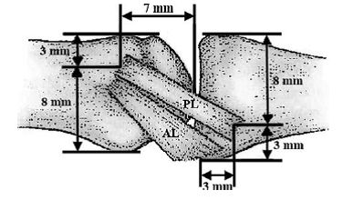
The UCL comprises of the proper ligament (PL) and the accessory ligament (AL). (Patel, Potty, et al (2010). Collateral Ligament Injuries of the Metacarpophalangeal Joint of the Thumb: A Treament Algorithm. Strat Traum Limb Recon . 5: 10)
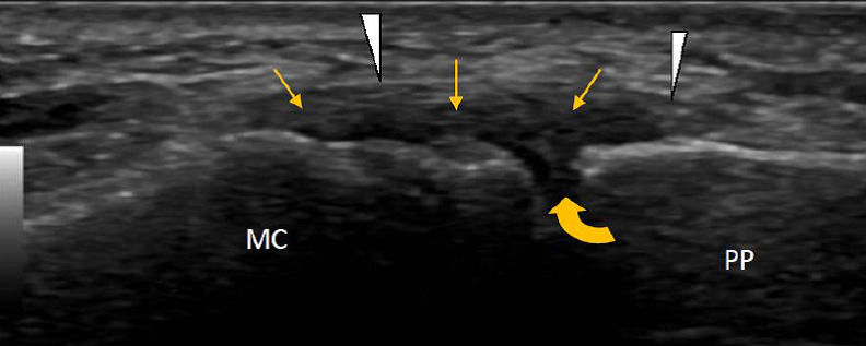
Longitudinal image of the UCL. The ligament appears as a low echo structure (straight yellow arrows) arching across the joint (curved yellow arrow). A thin anaechoic line maybe seen over the top of the ligament representing the adductor aponeurosis.
Testing should be carried out both in 30° flexion and neutral to assess both the ligament proper and accessory ligament respectively.
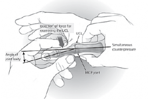
Injury to the UCL usually follows a forced abduction injury and may result in a partial tear (usually at the articular side of the ligament) or a complete tear. Both partial and complete tears may be associated with or without a bony avulsion.
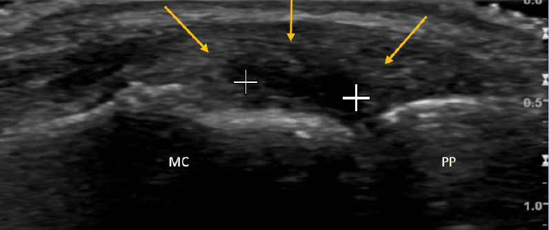
Longitudinal image of the UCL (yellow arrows). The ligament demonstrates marked thickening but also a low echo foci within its deeper aspect representing a partial articular side tear (white crosses).
A Stener lesion occurs when the adductor aponeurosis becomes interposed between the ruptured ends of the UCL. Such an interposition prevents healing and on US is seen as a
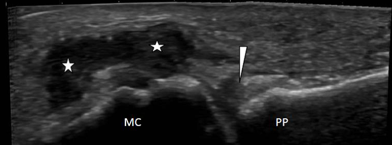
Longitudinal image of the UCL. The large anechoic lesion overlying the metacarpal head is in keeping with a Stener lesion. The distal stump of the ruptured UCL maybe seen over the base of the phalanx (white arrowhead).
The video clip demonstrates dynamic stressing of the UCL with a valgus force. The ligament appears thickened and there is an avulsion fracture noted to the left of the image. The avulsion fragment does however appear stable and the ligament patent with this dynamic test. (Click on link below for video)