US Case 3: Subluxation of the Long Head of Biceps
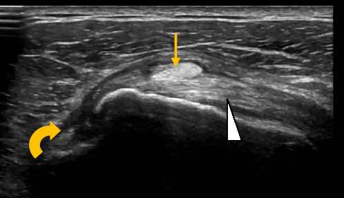
A 52 year old male patient presented to clinic with intermittent catching and pain at the anterior aspect of the shoulder following a press overhead with a barbell. An MRI had demonstrated a well sited long head of biceps within the bicipital groove. Likewise static US imaging demonstrated a well sited tendon. However, with active external rotation of the arm the tendon could be seen to sublux medially as demonstrated below.

This standard longtudinal image of the subscapularis tendon at its insertion onto the lesser tuberosity (white arrowhead) demonstrates the long head of biceps in transverse laying over the subscapularis (yellow arrow) having subluxed from the bicipital grove (to the left)

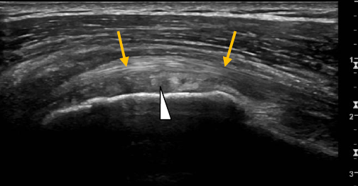
This transverse image of the subscapularis tendon at the lesser tuberosity (white arrowhead) demonstrates the long head of biceps (yellow arrows) laying superior.
