Case 14: Weber ‘A’ Fracture
A 40-year old female patient was referred for an US of the lateral ligaments to include dynamic assessment with a long history of ankle instability and pain. Interestingly she could recall no significant trauma although did describe a long history of relatively mild inversion injuries. No imaging had been undertaken to date. Physiotherapy had helped but she reported ongoing symptoms.
To scan the anterior talofibular ligament (ATFL) the patient should be positioned in supine with the leg internally rotated and the foot hanging off the couch. The probe is placed so that its posterior edge lies over the lateral malleolus and its anterior edge is over the lateral talus. The probe is almost parallel with the bottom of the foot.
This position allows the clinician to move the foot into a plantar fexed and inverted position placing a stress through the ATFL to assess dynamically for stability.
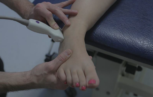
The US of the anterior talofibular ligament (ATFL) for this patient is shown below,
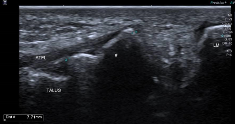
The lateral malleolus (LM) maybe seen to the right of the image. There is then a considerable gap before a bony fragment is seen. Attached to this fragment the ATFL appears intact and is seen to be attached distally to the lateral talus. Initially the probe was placed over the lateral malleolus and the bony fragment with the assumption that the ATFL was ruptured. However, moving the probe more distally demonstrated that the ATFL was intact. The problem being the large bony avulsion which on testing was very unstable in relation to the lateral malleolus.
Scanning the calcaneofibular ligament demonstrated similar findings and accounted for the significant instability described by the patient.
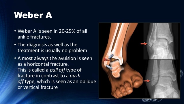
Correlating XR is shown below.
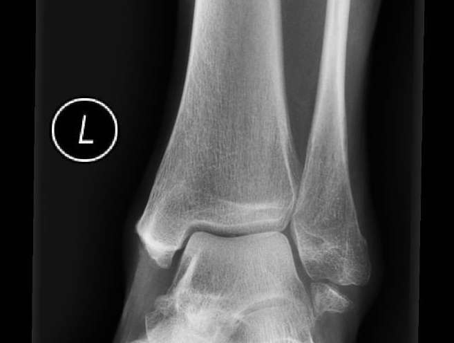
Findings are in keeping with a Weber-A type fracture.
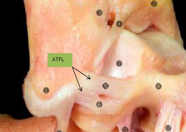
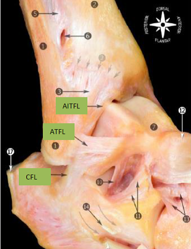
When scanning the lateral ligaments it is worth noting that the anterior inferior tibiofibular ligament (AITFL), anterior talofibular ligament (ATFL) and the calcaneofibular ligament (CFL) each run approximately 90 degrees to each other as below,
It is also worth noting that there are possible anatomical variations such as the ATFL being composed of 2 distinct bands as below,
