Case 12: Imaging the Distal Biceps Tendon
The traditional way to view the distal biceps tendon and the way I have used and taught has been to fully extend and supinate the elbow. In this position it is possible to see the tendon but imaging is often sub-optimal as the tendon ‘dives’ away from the probe to its insertion onto the tibial tuberosity.
This can be a particular issue if the patient presenting to clinic is suspected of having ruptured the tendon as they will not necessarily tolerate this position. The images below demonstrate this method and although the tendon can be seen it is relatively indistinct.
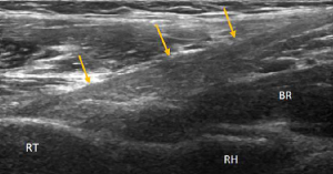
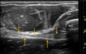
RH-radial head; RT-radial tuberosity; BR-brachialis; Yellow arrows- biceps tendon
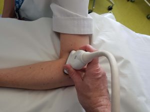
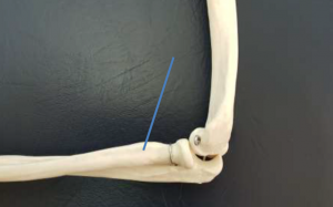
There are two possible alternative views. The first has the patient with the elbow bent to 90 degrees and the probe placed in line with the humerus. The probe is then moved forwards and angled slightly obliquely. The distal edge of the probe lays over the radius at the level of the radial tuberosity.

The image obtained maybe seen below. The probe views the distal biceps tendon (yellow arrows) through the overlaying brachioradialis muscle (BR). The two heads of supinator (SUP) maybe seen just to the right of the image overlaying the radius (Rad). Although the tendon maybe seen clearly the most distal insertion onto the radial tuberosity is still not well visualised as it lays on the deeper aspect of the radius.
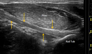
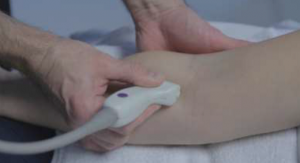
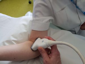
A better view is obtained from the medial aspect of the elbow which is again flexed to 90 degrees. The probe is placed in line with the humerus and then moved anteriorly to lay obliquely over the anteromedial elbow as demonstrated below.
The image produced (below) clearly demonstrates the distal bicep tendon (yellow arrows) deep to pronator teres (Pron) which lays in a transverse plane over the tendon. In this medial view the tendon maybe seen clearly down to its insertion onto the radial tuberosity.
Top tip: If you are struggling to find the tendon look for the brachial artery in the same plane and move the probe slightly anterior to this.