Case 11: Tear of the Fascia Cruris
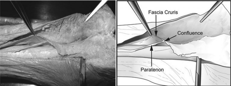
The fascia cruris is a layer of connective tissue which encloses the posterior structures of the calf extending as far down as the ankle joint with connection directly to the Achilles tendon. The paratenon which takes the place of a true synovial sheath surrounds the posterior, lateral and medial Achilles and lies deep to the fascia cruris.
Injury to the fascia cruris maybe diagnosed as an ‘atypical’ Achilles tendinopathy.
Ultrasound should be directed to the tender area with the probe in the transverse plane over the Achilles. Since the injury is usually to the medial or lateral side of the Achilles the probe should be appropriately orientated.
Injury to the fascia cruris is seen as a hypoechoic area which extends from either the medial or lateral Achilles in an anterior direction. Clinically as well as tenderness to direct palpation of the area there may also be some soft tissue swelling. Comparison with the contralateral side maybe useful. The hypoechoic region maybe associated with some increase in vascularity when Power Doppler is utilised.
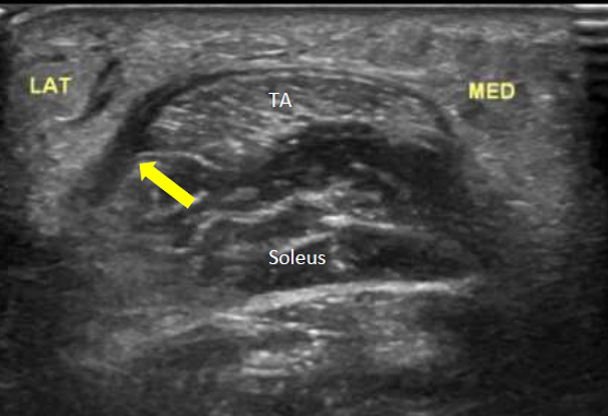
In the image below the probe has been placed in the transverse plane over the upper third of the Achilles tendon (TA). The Soleus muscle maybe seen deep to the Achilles. The yellow arrow points to a hypoechoic foci laying to the lateral side of the Achilles and extending anteriorly. The medial side appears normal. In this example the patient was a 44 year old runner who had felt a ‘pulling’ in his calf while taking part in some interval training 2 weeks prior to this scan.
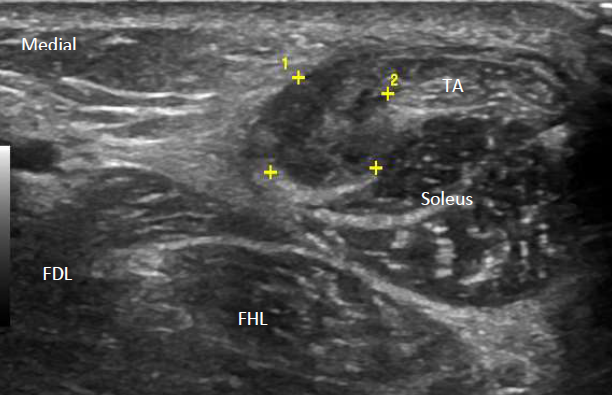
In another case below a relatively large hypoechoic region (yellow calipers) maybe seen extending around the medial side of the Achilles (TA) and overlying the Flexor digitorum longus (FDL) and Flexor hallucis longus (FHL) muscles.