
Case 10: Freiberg’s Disease
Freiberg’s Disease (also known as Freiberg’s infarction)
- Most commonly seen in 12 to 18 year old patients. However if missed it may become apparent when the joint develops secondary osteoarthritis later in life.
- Commonly occurs in the 2nd metatarsal head (approx. 70% of cases).
- Female to male ratio 4:1.
- Caused when there is a disruption of the blood supply leading to an avascular necrosis. The reason for this is not well understood.
Once established and painful guided injection may be a useful adjunct to a conservative management plan.
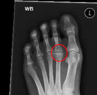
The XR demonstrates a loss of the normal rounded appearance of the 2nd metatarsal head with flattening and irregularity.
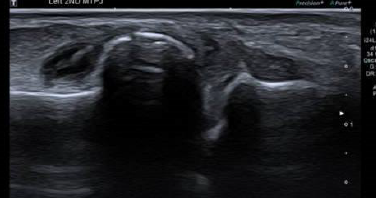
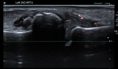
The longitudinal US images demonstrate the cortical irregularity of the metatarsal head demonstrated in the XR. The base of the proximal phalanx to the left of the image appears relatively well preserved. In addition to the cortical change US demonstrates clear soft tissue hypertrophy, fluid and some Doppler signal all in keeping with synovitis of the joint.
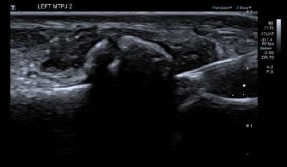
Injection is given from a distal to proximal direction in plane with the probe and the needle aimed at the more prominent metatarsal head. This approach would be utilised for injection of any of the metatarsophalangeal joints.