
US Case 1: Anatomical variations in Dorsal Compartment 1 (DC1) of the Wrist
The tendon of abductor pollicis longus (APL) and extensor pollicis brevis (EPB) lie side by side within the first dorsal compartment of the wrist lateral to the radial styloid. APL is in the more volar position with EPB laying more dorsally.
A possible anatomical variant often seen with US examination of the wrist is a central vertical septum separating the APL and EPB tendons as demonstrated below. This central septum maybe seen to form a vertical hypoechoic band. The volar part of the tunnel contains the larger APL and the dorsal part the smaller EPB.
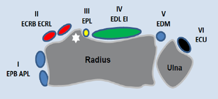
The 1st DC maybe seen to be the post lateral of the DC’s
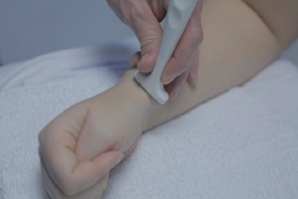
The patient places their forearm in a neutral position with the thumb uppermost.
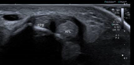
Transverse image of DC1 of the wrist. APL maybe seen laying to the right of EPB. A hypoechoic band is seen running vertically between the two tendons in keeping with an internal septum.
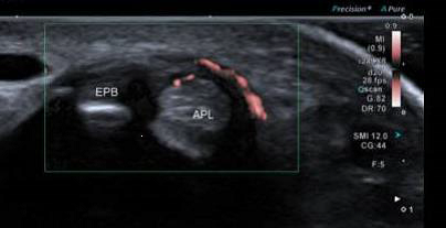
In this image Doppler demonstrates that there is an increased vascularity within DC1 but only around the tendon of APL which is separated from EPB by an internal septa.
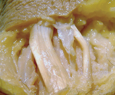
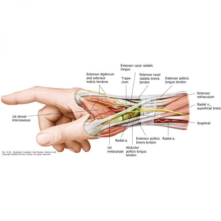
The tendon of APL is to the left of the image and EPB to the right. The white tissue between the two tendons corresponds to the hypoechoic internal septa seen on US.
The other possible variant would be when APL splits into multiple slips with in DC1 giving the appearance of 4 or 5 tendons within the tunnel. Anyone have a good image of this?